AUSTIN, Texas—Scientists have learned how to create protein barriers near living nerve cells that influence their direction of growth, which could one day provide a way to precisely control nerve-cell interactions to better understand memory formation and other brain functions.
“To get complex brain behavior, nerve cells need to integrate information they are receiving from other nerve cells in a very complex way that depends partly on where along the cell it physically contacts those signaling nerve cells,” said Jason Shear, the lead researcher from The University of Texas at Austin.
“It would be a powerful thing if we had the capacity to control where a particular contact is made.”
In a paper published online Monday (Nov. 8) by the Proceedings of the National Academy of Sciences, Shear’s laboratory demonstrated that they could build microscopic walls of protein near a nerve cell growing on a glass slide after a thick liquid containing dissolved copies of the protein was added.
The microscopic walls developed by forming chemical bonds, or cross-links, between copies of the protein to produce a dense interconnected network. Another chemical in the soupy liquid known as a photosensitizer was activated by laser light to generate reactive chemicals that drove the proteins’ interaction. Shear and his laboratory group were able to identify photosensitizers such as flavin adenine dinucleotide that were effective without being toxic to cells.
“A major part of this effort was finding photosensitizers that worked well, but also were compatible with cells,” he said.
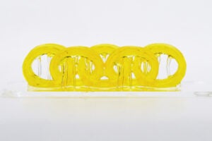
Using this laser-induced process of building protein structures called microfabrication, the group built walls near living nerve cells that were made of albumin or other proteins. Shear’s laboratory also demonstrated that a wall could be used to guide and direct two nerve cells to interact at a specific site at the end of a wall.
The titanium/sapphire laser used in the experiments allowed for the precision needed. These lasers produce short pulses of light that meant Shear’s group could produce tiny spots of clumped proteins as close as 1.5 microns (0.0015 millimeters) to a cell without inducing any apparent cell damage.
The laser’s position also could be adjusted quickly enough to extend walls in milliseconds. In unpublished experiments, for instance, Shear’s laboratory was able to momentarily trap bacteria inside a protein “room” before they crawled over one of its walls.
These bacterial experiments also were important for testing another potential use of protein microfabrification: the tethering of biologically important molecules onto a wall that cells are growing near. In the case of nerve cells, molecules called chemoattractants that lure in the cells could be tethered to sites where a scientist wants two nerve cells to interact.
Shear and his lab demonstrated that they could tether a similar attractant molecule to a protein wall in a container of bacteria and have them rapidly migrate to that site.
“We want to make this microfabrication guidance method capable of creating a much more complex nerve-cell landscape—not just physical barriers, but ones that also interact with cells chemically and electronically,” he said.
Note: Graduate students Bryan Kaehr and Richard Allen were among those conducting the studies for the paper.
To obtain an online copy of the article “Guiding neuronal development with in situ microfabrication,” visit the Proceedings of the National Academy of Sciences Web site. For a black-and-white image of a nerve cell whose outgrowth (neurite) was forced to grow along a protein wall, contact Barbra Rodriguez at brodriguez@mail.utexas.edu.
For more information contact: Barbra Rodriguez, College of Natural Sciences, 512-232-0675.


