Do you remember the first time you saw a microscopic image of a fly? Those huge eyes, the tiny hairs, the delicate wings? That monstrous picture was possible thanks to a simple microscope, a revolutionary tool invented in the 17th century.
Although giant flies may have been a little startling, you were probably accustomed to the wonders of 20th century technology. Imagine the fascination or horror experienced by readers in 1665 of Robert Hooke’s book, Micrographia: Or Some Physiological Descriptions of Minute Bodies, which brought to life the fantastic miniature world revealed by the new technology.
In the Micrographia’s original illustrations, now canonical in the history of science, readers saw for the first time magnified details of ordinary objects, including lice, flies, seed pods, a razor’s edge and a needle’s blunt-looking tip. With these images, Hooke’s book the first science bestseller revolutionized ways of seeing even for non-scientists.
Now, University of Texas at Austin researchers are focusing modern microscopy and visualization technologies on that very book. This joint project of the Department of English and the Texas Advanced Computing Center, with help from the Institute for Cell and Molecular Biology (ICMB), shows the essence of a physical book at the microscopic and atomic levels.
The project uses two modern microscopy techniques to interrogate this famous text.
First, the team is creating a microscopic panorama of a single page of Micrographia (from a 1667 second-edition copy) by stitching together hundreds of photos taken with a stereo dissection microscope in the genetics lab of Janice Fischer, a professor in Molecular Cell and Developmental Biology.
The photos were painstakingly shot one by one, moving the microscope’s camera across the page in millimeter increments. These photos were then stitched together into an integrated image using the expertise and processing software at the TACC/ACES Visualization Laboratory (Vislab).
Digitally suturing the images, or tiles, together makes it possible to create a single image of the entire page that contains an incredible amount of microscopic detail. However, because the final image is so large, only a tiny portion of the image would be viewable on a standard computer monitor. To view the image in its full glory, the researchers loaded it on the 36-foot-wide “Stallion” display wall at the TACC/ACES Vislab.
Made up of 80 individual monitors, Stallion boasts a whopping 328-megapixel resolution, making it the highest-resolution-tiled display in the world. Seen in this way, the team’s images of Hooke’s book have the same blow-your-mind-wow-factor that Hooke’s oversized engravings must have had in the 1660s.
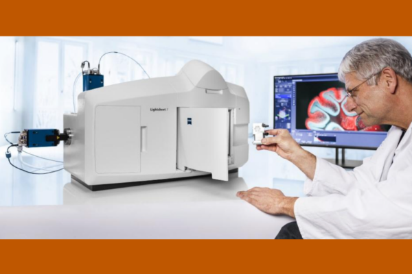
Finally, to get an even “deeper” perspective on the book, the team turned to scanning electron microscope technology, which can create images down to the atomic level. Electron Microscopy Specialist Dwight Romanovicz ran samples taken from the Micrographia‘s pages through the Zeiss Supra 40 VP Scanning Electron Microscope, a facility of the ICMB. The SEM image takes the microscopic view further than Hooke, who coined the term “cell,” could have imagined in the 17th century and does so using his own work as the subject.
“Turning advanced microscopy back on the book that started the field has been an amazing experience,” says Janine Barchas, associate professor of English and co-director of “The Fate of the Book” symposia. “When you’re allowed to use the tools and expertise of colleagues from completely different research areas, you see things in a new light.”
The Micrographia project was conceived to kick off the Department of English’s yearlong “The Fate of the Book” symposia, sponsored by the Texas Institute for Literary and Textual Studies (TILTS).
Elizabeth Scala, associate professor of English and symposia co-director, says “Hooke’s Micrographia is not just a convenient historical example of a comparable pivotal technology, but a fitting object to digitize in order to ask at what point digitization changes a book, or its text, into something else.
“Digital publishing technology affects everyone who reads, regardless of discipline,” Scala says.
A “Preservers Panel” discussion on Oct. 25 at the Harry Ransom Center is the next event in “The Fate of the Book” symposia series. See the full schedule of the series, which continues through the spring.
Note: Images of the high-resolution Micrographia scans will be on view at the TACC/ACES Visualization Laboratory by appointment.