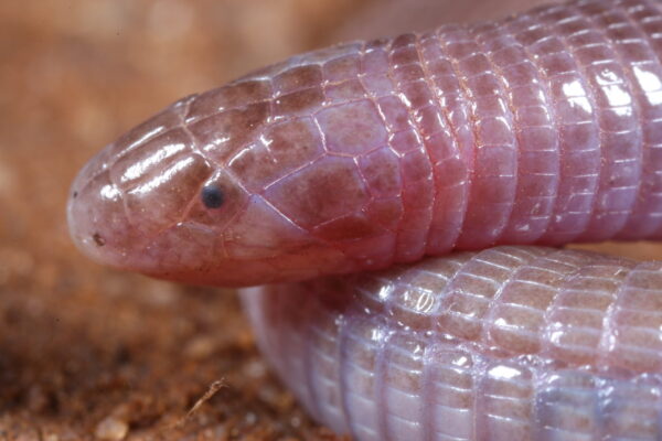
AUSTIN, Texas — Amphisbaenians are strange creatures. Like worms with vertebrae, scales, a large central tooth and sometimes small forearms, these reptiles live underground, burrowing tunnels and preying on just about anything they encounter, not unlike a miniature version of the monstrous sandworms from “Dune.”
Even though they’re found around much of the world, little is known about how amphisbaenians behave in the wild because they cannot be observed while in their natural habitat under sand and soil. But thanks to two papers published in the March issue of The Anatomical Record, new light is being shed on these animals and their specialized anatomy.
Using a micro-CT scanner at The University of Texas at Austin’s Jackson School of Geosciences, researchers completed a detailed comparative analysis of 15 amphisbaenians from southern Africa and a bone-by-bone description of every cranial anatomical feature of the species Zygaspis quadrifrons. These are the most detailed studies of southern African amphisbaenians to date, according to the researchers.
By CT scanning these specimens, researchers were able to render individual bones as large 3D-printed models. This made it possible for them to take a close look at bones such as the tabulosphenoid, which is almost entirely inside the skull and almost impossible to see without this technology, said Christopher J. Bell, the lead author on the paper that delved into the skull anatomy of Zygaspis, and a professor in the Jackson School.
“You could fit three skulls of the Zygaspis quadrifrons on the nail of my pinky. We can now look at these really small vertebrate organisms in a measure of detail that we never had before,” Bell said.

The research began more than 15 years ago when Patrick J. Lewis, a co-author on both papers and paleobiology professor at Sam Houston State University, led a team to Botswana on a mission to trap and study animals of all kinds. While digging and sampling the environment, they began catching amphisbaenians. At the time, Lewis didn’t know much about them. When one of his students handed him one, he said he was surprised that something that looked so much like a worm could be so strong.
“They wriggle around and try to escape and move in ways that worms just aren’t able to. These are much more like little snakes in the way that they move and interact. It’s just surprising for something that’s so tiny. You just don’t expect that behavior,” Lewis said.
Some of the most striking imagery to come from these CT scans highlights sutures within the skull: deep, thin waves that “grab” on to each other, Lewis described. The images also render in exquisite details the amphisbaenians’ strange singular central tooth, which interlocks with two bottom teeth.
“Combined with the powerful jaw muscles in amphisbaenians, it gives them a ferocious bite for an animal of their size. They can bite and tear out pieces of their prey,” Bell said.
Antonio Meza, a lead author on the paper examining the different species of amphisbaenians and first-year doctoral student at Arizona State University, said that snakes and other reptiles are born with an egg tooth that allows them to break out of their shell.
“But in amphisbaenians, they just have kept it,” he said.

Meza’s analysis also confirmed sexual dimorphism in Zygaspis quadrifrons, with the females larger than males in this species.
With little biological and ecological data available on amphisbaenians, studying their anatomy is the best way for researchers to learn more about these bizarre animals and the hidden lives they lead beneath the surface.
The research was funded by the National Science Foundation, the Jackson School of Geosciences and Sam Houston State University.